
“Hyperdense Mucus Plug” Sign – CT Findings In Allergic Bronchopulmonary Aspergillosis
DOI:
https://doi.org/10.56412/gmcp.2026.9.01.219Keywords:
Allergic Bronchopulmonary Aspergillosis (ABPA), High-resolution Computed tomography (HRCT), High-attenuation mucus (HAM)Abstract
Background: Allergic Bronchopulmonary Aspergillosis (ABPA) is a hypersensitivity response to Aspergillus fumigatus with varied radiological manifestations. While the differential diagnosis of central bronchiectasis is broad, identifying pathognomonic radiological features on high-resolution computed tomography (HRCT) is essential for an accurate diagnosis.
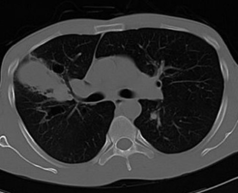
Case Presentation: A 52-year-old female with a history of asthma presented with a chronic cough and expectoration. Laboratory workup demonstrated elevated eosinophil counts and total serum IgE. Chest radiography revealed central bronchiectasis with a right upper lobe radio-opaque shadow. Unenhanced HRCT confirmed bilateral central bronchiectasis and identified hyperdense mucus plugging in the right upper lobe bronchi (density = 101 HU) distal to a large bronchocele, establishing a diagnosis of ABPA with high-attenuation mucus (ABPA-HAM). The patient was successfully treated with systemic corticosteroids and oral antifungals.
Discussion: High-attenuation mucus (HAM) is defined as mucoid impaction denser than normal paraspinal skeletal muscle. Present in up to 30% of ABPA patients, HAM correlates with increased disease severity, higher baseline serum IgE, and a greater risk of relapse following therapy.
Conclusion: The "hyperdense mucus plug" sign is a characteristic and virtually diagnostic HRCT finding in ABPA. Early recognition is critical for prompt therapeutic intervention and long-term prognostic monitoring.
Downloads
References
1. Webb WR, Müller NL, Naidich DP. High-Resolution CT of the Lung. 4th ed. Philadelphia: Lippincott Williams & Wilkins; 2009.
2. Agarwal R, Aggarwal AN, Gupta D. High-attenuation mucus in allergic bronchopulmonary aspergillosis: another cause of diffuse high-attenuation pulmonary abnormality. AJR Am J Roentgenol. 2006;186(3):904. doi: 10.2214/AJR.05.0125
3. Agarwal R, Gupta D, Aggarwal AN, Saxena AK, Chakrabarti A, Jindal SK. Clinical significance of hyperattenuating mucoid impaction in allergic bronchopulmonary aspergillosis: an analysis of 155 patients. Chest. 2007;132(4):1183-1190. doi: 10.1378/chest.07-0808
4. Agarwal R, Gupta D, Aggarwal AN, Saxena AK, Saikia B, Chakrabarti A, Jindal SK. Clinical significance of decline in serum IgE levels in allergic bronchopulmonary aspergillosis. Respir Med. 2010;104(2):204-210. doi: 10.1016/j.rmed.2009.09.005
5. Silva CIS, Colby TV, Müller NL. Asthma and associated conditions: high resolution CT and pathological findings. AJR Am J Roentgenol. 2004;183(3):817-824. doi: 10.2214/ajr.183.3.1830817
6. Goyal R, White CS, Templeton PA, et al. Case report. HAM plugs in ABPA: CT appearance. J Comput Assist Tomogr. 1992;16(4):649-650. doi: 10.1097/00004728-199207000-00028
7. Logan PM, Primack SL, Müller NL, et al. Invasive aspergillosis of the airways: radiographic, CT, and pathological findings. Radiology. 1994;192(3):383-388. doi: 10.1148/radiology.193.2.7933881
8. Patterson TF, Thompson GR, Denning DW, Fishman JA, Hadley Susan, Herbrecht R, et al. Practice guidelines for the diagnosis and management of aspergillosis: 2016 update by the Infectious Diseases Society of America. Clin Infect Dis. 2016 Aug 15;63(4):e1-e60. doi: 10.1093/cid/ciw326.
Published
How to Cite
Issue
Section
License
Copyright (c) 2026 Rekhi AK, Brar RS, Dwivedi A, Deepander S Rathore, Puri S, Aggarwal V, Rana N, Sandhu K, Rana R

This work is licensed under a Creative Commons Attribution 4.0 International License.


